Microscopy Facilities
The Core provides access to a range of microscopy systems and ultra-high specification image analysis workstations, details of these systems are shown below
 |
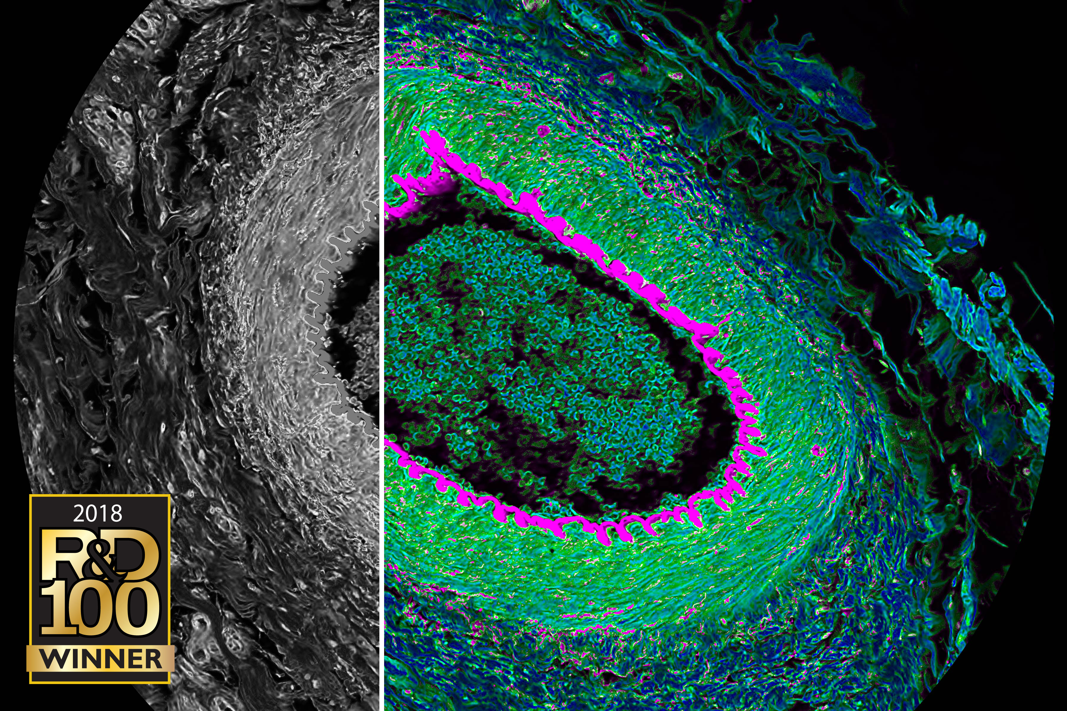
The Zeiss LSM900 is a point scanning confocal microscope, which in combination with the second generation Airyscan detector allows user friendly super resolution imaging without the need for complex sample preparation protocols. This system includes a fully motorised stage with piezo z drive for rapid acquisition of multiple positions, with the option to z stack and tile positions seamlessly. This system is also equipped with full incubation for live cell experiments including temperature, humidity and co2 control. In combination with the incredibly sensitive Airyscan detector, this allows long timelapse super-res imaging of cellular dynamics with minimal impact on cell viability. Excitation - diode lasers
Objectives
Software;
|
 |
Olympus SpinSR SoRa with high content imaging The Olympus SpinSR SoRa is a spinning disc confocal microscope equipped with software for both standard microscopy experiments and high content multiwell plate assays. The Yokogawa CSU-W1 SoRa spinning disc unit features both a 50um pinhole disc for confocal imaging, and a SoRa super-res disc, allowing you to switch imaging modalities at the touch of a button. This enables rapid acquisition of large areas of interest, even when including z stacking. The system is very sensitive due to the camera based detection. This in combination with full co2, humidity and temperature controlled environment allows rapid acquisition of dynamic events, whilst maintaining long term viability. The microscope is also equipped with DIC for high contrast brightfield acquisition. Excitation - diode lasers
Objectives
Detection
Software
|
 |
The SP8 Fast Lifetime Contrast (FALCON) is a point scanning confocal microscope with lifetime imaging and analysis built in to the acquisition software. The system has a fully automated stage and 7 fixed laser lines, allowing fast acquisition of multichannel, multi-position experiments. The microscope also features a pulsed 440nm laser for lifetime experiments. This in combination with a high sensitivity single photon time-gated counting spectral detection array and full temperature, humidity and co2 incubation allows a diverse range of experiments to be performed on the microscope. Excitation
Objectives
Software
|
 |
The Leica SP8 is a point scanning confocal microscope which features a white light laser for excitation. This allows selection of any excitation wavelength within the range of 470-670nm, which in combination with the standard fixed lines on the system allows careful excitation of almost all commonly used fluorophores. The SP8 also features a unique spectral detection array, allowing careful separation of emission from multiplex assays. Excitation
Objectives
Detection
Software
|
 |
Leica DMI8 Widfield (Ca+ imager) The Leica DMI8 is a widefield microscope equipped with both a monochrome camera for high sensitivity fluorescence assays, and a colour camera for histopathological stains. The system features an automated stage which in combination with the navigation and multi-well plate modules in the software and fast filter wheels allows rapid acquisition of large fields of your sample in multiple channels. The dual camera setup is ideal for protocols requiring colorimetric stains on tissue sections to give spatial context to fluorescent probes. Excitation
Objectives
Software
|
 |
The EVOS M5000 is a widefield microscope available to image fluorescent and colorimetric samples within our lab space. The system features a very simple user interface which is ideal for checking staining’s and capturing basic widefield fluorescent images. The microscope comes equipped with long working distance lenses with phase contrast capabilities, and therefore does not require that samples be placed in to imaging plates or coverslips. Excitation
Objectives – long working distance;
Detection
Software
|
 |
The Elyra PS1 is a widefield fluorescent microscope which combines TIRF and SMLM modalities in to a single system. Single molecule localisation microscopy uses the phenomenon of fluorophore ‘blinking’ to push the resolution limit of light microscopy down to the 30-40nm scale. TIRF imaging allows high resolution imaging of structures close to the coverslip with fantastic signal to noise ratio, and so is ideal for imaging structural proteins and targets which sit on the cell surface. This system features a dual camera setup, allowing rapid simultaneous multichannel acquisition. Excitation
Objectives
Detection
Software
|
 |
Zeiss Microbeam Laser Capture Microscope The Zeiss Microbeam Laser Capture Microdissection offers unique ‘laser catapulting’ technology. This involves using a high power UV laser to cut around regions of interest on tissue sections, down to the single cell level. The system then uses a defocused pulse of laser light to catapult the cut area in to the lid of a tube, which can then be taken for downstream analysis. The microscope comes equipped with a robomover automated sample capture system, which allows cuttings to be automatically directed to up to 96 different caps within a single experiment. Samples can be imaged and cut out using both colorimetric and fluorescent protocols. Fluorescence
Objectives
Detection
|
The Cellular Imaging Core is located on the ground floor, Laboratory 1, in room's 00/070, 00/071, 00/072 and 00/075. Only Core trained users have access to the Core microscopes. The microscopes can be used by all Wellcome Trust Centre staff and visitors once they have been trained and registered by Core staff. Please see the cost recovery page for hourly recharge rates.
Email us for further details or for new user microscopy induction at cellular-imaging@well.ox.ac.uk. Telephone 01865 287568 (internal ext. 87568) for the Core laboratory 00/069. You must be on the Wellcome Trust Centre's intra-net for some links to work.
